
I was born with a congenital heart defect called pulmonary atresia with intact ventricular septum and moderate hypoplasia of the right ventricle. The information below the "My Heart Timeline" describes my congenital heart defect. My Mommy and Daddy can give you more information if you want.
My Heart Timeline:
July 12, 2005 – Bang, zoom, whoosh, I'm here. Let's rock. At around 5 hours old, the nurses noticed that I was a little cyanotic (blue). I was sent to the NICU for observation and then transferred to the Infant Intensive Care Unit at Children's Hospital. The next day I was diagnosed with a congenital heart defect called pulmonary atresia with intact ventricular septum and moderate hypoplasia of the right ventricle.
July 14, 2005 (2 days old) – I had radiofrequency valvotomy and balloon dilation of the right ventricle followed by a patent ductus ateriosus stent insertion via cardiac catheterization. The goal is to eventually establish complete biventricluar circulation. I was released from Children's Hospital when I was 6 days old.
October 20, 2005 (3 months old) – Cardiac catheterization was once again performed by Dr. Jones at Children's Hospital. This time it was a balloon valvoplasty on my right ventricle and PDA stent. This procedure was needed to improve blood flow through my pulmonary valve and my PDA. I was released after an overnight stay.
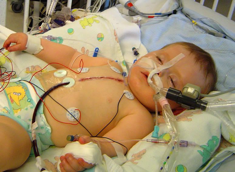
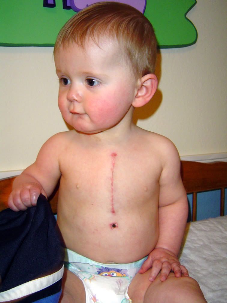 April 20, 2006 (9 months old) – Open heart surgery at Children's Hospital was performed by Dr. Permut. The easy name for this surgery is called right ventricle outflow tract transannular patch surgery. The patch was taken from the pericardium (heart lining) of a cow. The pediatric cardiac surgeon made a vertical incision over my pulmonary valve, spread the tissue apart and stitched the patch over that area. The goal was to create sufficient blood flow through the ventricle and into the lungs. I was on the heart/lung bypass machine during the surgery. After the surgery, I spent several days in the cardiac ICU. They had to use some powerful medication to keep me from moving around and messing with the tubes and wires that were attched to my chest. I also had some fluid around my lungs so I had to continue using the ventillator until it cleared. On April 24, I was freed from the ventillator and only connected to an EKG and a blood saturation monitor. April 25, I am in a regular room, free of all wires, and ready to go home, but I am having problems keeping food down. I have an ear infection, a tooth coming in and rotavirus which explains my gastrointestinal issues. May 2, released from hospital after successfully joining the "zipper club."
June 2, 2010 (4 years, 11 months old) – I have an atrial septal defect (hole in heart between the two upper chambers) that needs to be closed via cardiac catheterization. During this procedure, an Amplatzer Septal Occluder is delivered via catheter through an incision in my groin. From there, it is moved through blood vessels until it reaches my heart. It expands to place a disc on either side of the heart defect, closing the hole between the left and right atria. I should be able to go home after an overnight stay in the hospital.
February 3, 2011 (5 years, 7 months old) – Not heart related...I had my appendix removed.
Future – Lifelong appointments with cardiologist to monitor my heart. Pulmonary valve replacement through either surgery or catheterization when needed.
- - - - - - - - - - - - - - - - - - - - - - -
The below information was copied from a medical website so the language is pretty dry and it doesn't exactly describe my case since all heart defects are anatomically unique. Given that disclaimer, it does go over the basics to give you a little general information.
 Pulmonary Atresia with Intact Ventricular Septum (PA / IVS)
Pulmonary atresia with intact ventricular septum (PA/IVS) is a rare congenital heart disease. Pulmonary atresia is when the valve that lets blood flow from the lower-right chamber (right ventricle) of the heart to the lungs has not formed properly or is closed. This abnormality is present from the very early stages of heart development (around the first eight weeks of pregnancy), resulting in an underdeveloped right ventricle, which is the pumping chamber of the heart.
Before birth, while the baby is developing, this condition does not threaten life because the placenta provides oxygen for the baby and the lungs are not yet functioning. Blood entering the right side of the fetal heart passes through an opening called the foramen ovale that allows oxygen-rich (red) blood to pass through to the left side of the heart and circulate throughout the body. After birth, the placenta no longer provides oxygen for the newborn — the lungs must provide it.
Two connections within a newborn's heart can provide temporary help in getting blood flow to the lungs — the foramen ovale and the patent ductus arteriosus (PDA). As these connections spontaneously begin to close after birth, the baby becomes critically ill due to an inadequate blood flow to the lungs where the blood picks up oxygen. This situation cannot support life, since oxygen-poor (blue) blood cannot meet the body's demands. The baby becomes very blue (cyanotic) and sick.
Receiving a diagnosis of PA/IVS can be frightening and overwhelming; however, there are surgical options for treating infants with this condition.
Diagnosis
Upon diagnosis, the baby is admitted to an intensive care unit. The baby may be treated with oxygen, a ventilator to assist breathing and intravenous medications to help the heart and lungs function more efficiently. In addition, the drug prostaglandin is administered to prevent the ductus arteriosus from closing, thereby maintaining blood flow to the lungs. This helps stabilize the baby while subsequent treatment decisions are made.
Treatment
Once stabilized, there are surgical treatment options:
Cardiac Catheterization
This procedure can be used as an initial treatment and a diagnostic tool for some cases of PA/IVS. A cardiac catheterization usually will be performed to evaluate any heart defects, establish whether the foramen ovale or ductus arteriosus are still open, and determine the amount and proportions of oxygen-rich (red) and oxygen-poor (blue) blood.
Balloon Atrial Septostomy
This procedure may be performed as part of the cardiac catheterization to improve the proportions of red blood and blue blood between the right and left atria (upper chambers of the heart). A special catheter (tube) with a balloon in the tip is used to create an opening in the atrial septum (the wall between the left and right atria). The catheter is guided through the foramen ovale and into the left atrium, and the balloon is inflated.
Radiofrequency Perforation
Radiofrequency Perforation of the pulmonary valve: In some cases it may be possible to burn a way through the blocked pulmonary artery. A fine tube is inserted into a vein and passed through the heart. A hot wire is used to burn into the pulmonary artery. A balloon is then inserted and used to stretch the pulmonary valve and artery. This does not leave any scar.
Open-heart Surgery
Ultimately, surgery will be necessary to improve blood flow to the lungs on a permanent basis. The types of operations for pulmonary atresia varies quite widely depending on the individual child’s cardiac anatomy and condition. A series of operations may be necessary to bypass the blocked pulmonary artery. These are open heart surgeries - the heart will need to be stopped and opened to repair it. This means that a machine will have to take over the job that the heart normally does – the heart bypass machine.
Following surgery, the baby's circulation may be normal. However, patients receiving a valve replacement may need another heart valve replacement surgery within the next 10 years as the new valve slowly wears out. The right-side pumping chamber (ventricle) is typically smaller than normal, so the baby may require another mechanism for blood flow to reach the lungs so enough oxygen can get to the body. In such a case, an artificial connection (shunt) between the aorta — the large artery that leaves the heart and provides oxygen-rich blood to the body — and the pulmonary artery is created to deliver blood to the lungs.
Additional Heart Surgery
Additional surgery is needed when the child is older, particularly in cases where the right ventricle was very underdeveloped. The kind of surgery recommended depends on the size of the right ventricle and the pulmonary artery. If they are normal size and the right ventricle is able to pump blood, open-heart surgery will make blood flow through the heart in a normal pattern. If the right ventricle is small and unable to pump, doctors may perform an operation called the Fontan procedure, in which the right atrium is connected directly to the pulmonary artery.
Follow Up
Children who have PA/IVS require intensive monitoring both before and after surgery, including blood tests, X-rays and urine tests. The child will also need lifelong monitoring by a congenital heart specialist.
Patients born with PA/IVS have a risk of developing endocarditis, an infection in the heart's walls or valves. Consequently, they require antibiotics before dental work and certain surgeries to prevent endocarditis.
No comments:
Post a Comment